
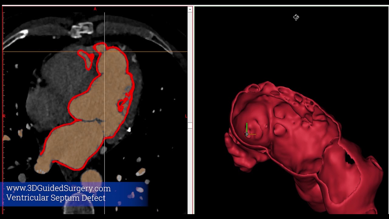
Following a large cardiac infarction, this man had developed a large hole between both cardiac cavities (a so-called ‘Ventricular septum defect’) for which he underwent open cardiac surgery. In follow-up echocardiography a remaining leak of 10mm was noticed. 3DGuidedSurgery informed the cardiologist and cardiac surgeon on the required imaging sequence and developed a 3D model to evaluate the leakage and measure it up nicely as this was also difficult to evaluate for the radiologist.
Next the model was also 3D printed to discuss percutaneous versus open versus conservative treatment. The model allowed for pre-operative fitting of the endovascular device.

simple text ###
Your consent is required to display this content from youtube - Privacy Settings
GET IN TOUCH
Have an idea or a project, lets collaborate!